Bone health is a critical aspect of overall wellness and quality of life. It’s a complex interplay of genetics, nutrition, lifestyle, and environmental factors. As our understanding deepens, so do the tools we use to assess and monitor the health of our bones. Recent advances in bone density imaging techniques are pivotal in the early detection and management of diseases like osteoporosis, and also in evaluating the effectiveness of treatments and lifestyle modifications aimed at improving bone health.
Understanding Bone Density and Its Importance
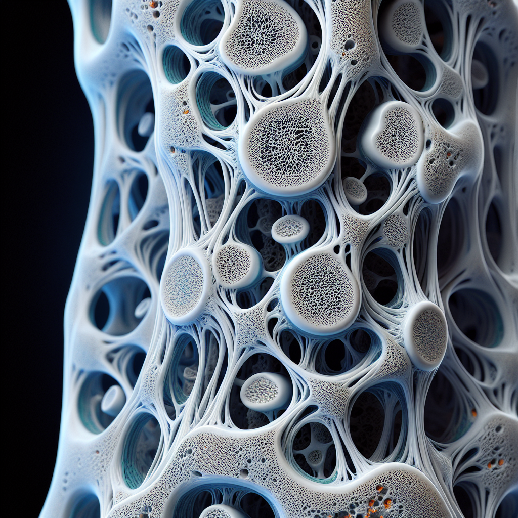
Bone density refers to the amount of mineral matter per square centimeter of bones. It is a crucial indicator of bone strength and an important diagnostic tool for assessing the risk of fracture. While a decrease in bone density is a natural part of aging, certain conditions and lifestyle factors can accelerate this process, leading to fragile bones that are more susceptible to breaks and fractures.
One of the most relevant aspects of maintaining bone health is understanding the effects of aging on bone structure. As we age, our bones can lose density and strength, making knowledge of one’s bone health increasingly important with time.
Evolution of Bone Density Imaging
Traditional imaging techniques, such as dual-energy X-ray absorptiometry (DXA), have long been the standard for measuring bone mineral density (BMD). DXA scans are quick, painless, and provide detailed information about bone density in various parts of the body. However, these scans have limitations, including the use of ionizing radiation and the inability to capture bone quality — a factor just as critical as bone quantity.
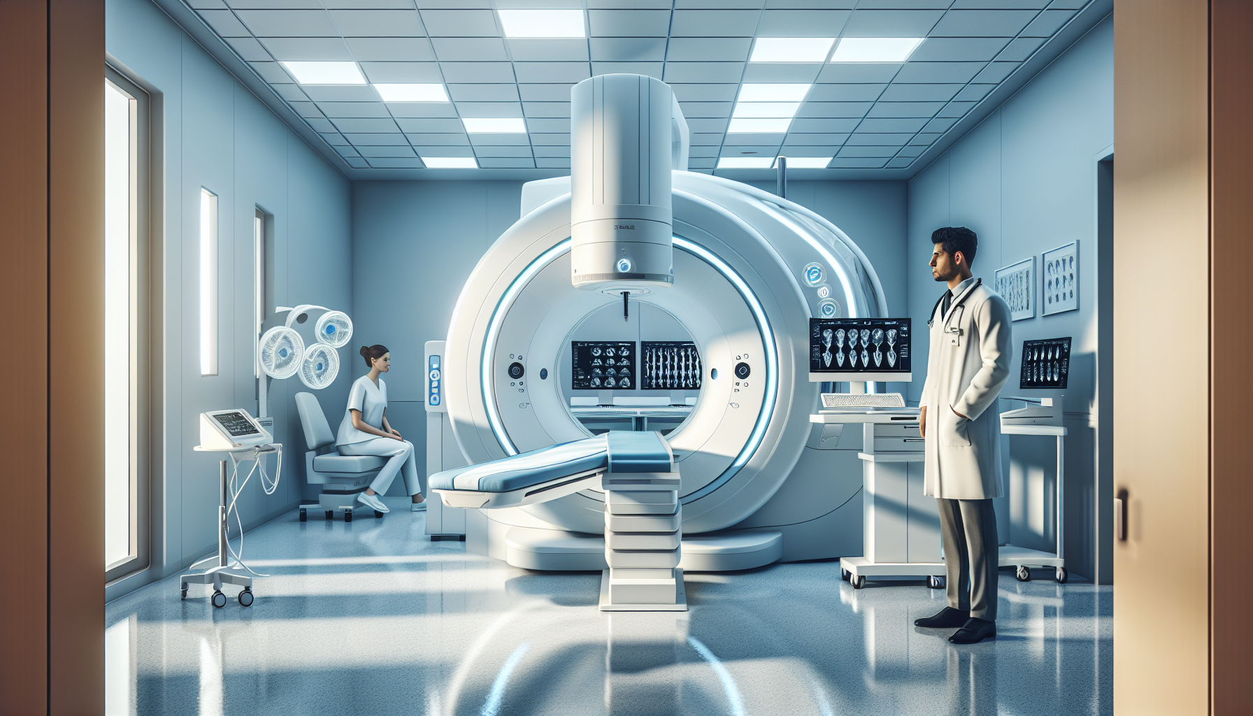
Enter the new era of bone density imaging, introducing sophisticated methods like high-resolution peripheral quantitative computed tomography (HR-pQCT) and magnetic resonance imaging (MRI). These techniques not only measure bone density but also provide intricate details about bone architecture and strength, offering a more comprehensive view of bone health.
Cutting-Edge Techniques in Bone Density Imaging
High-Resolution Peripheral Quantitative Computed Tomography (HR-pQCT)
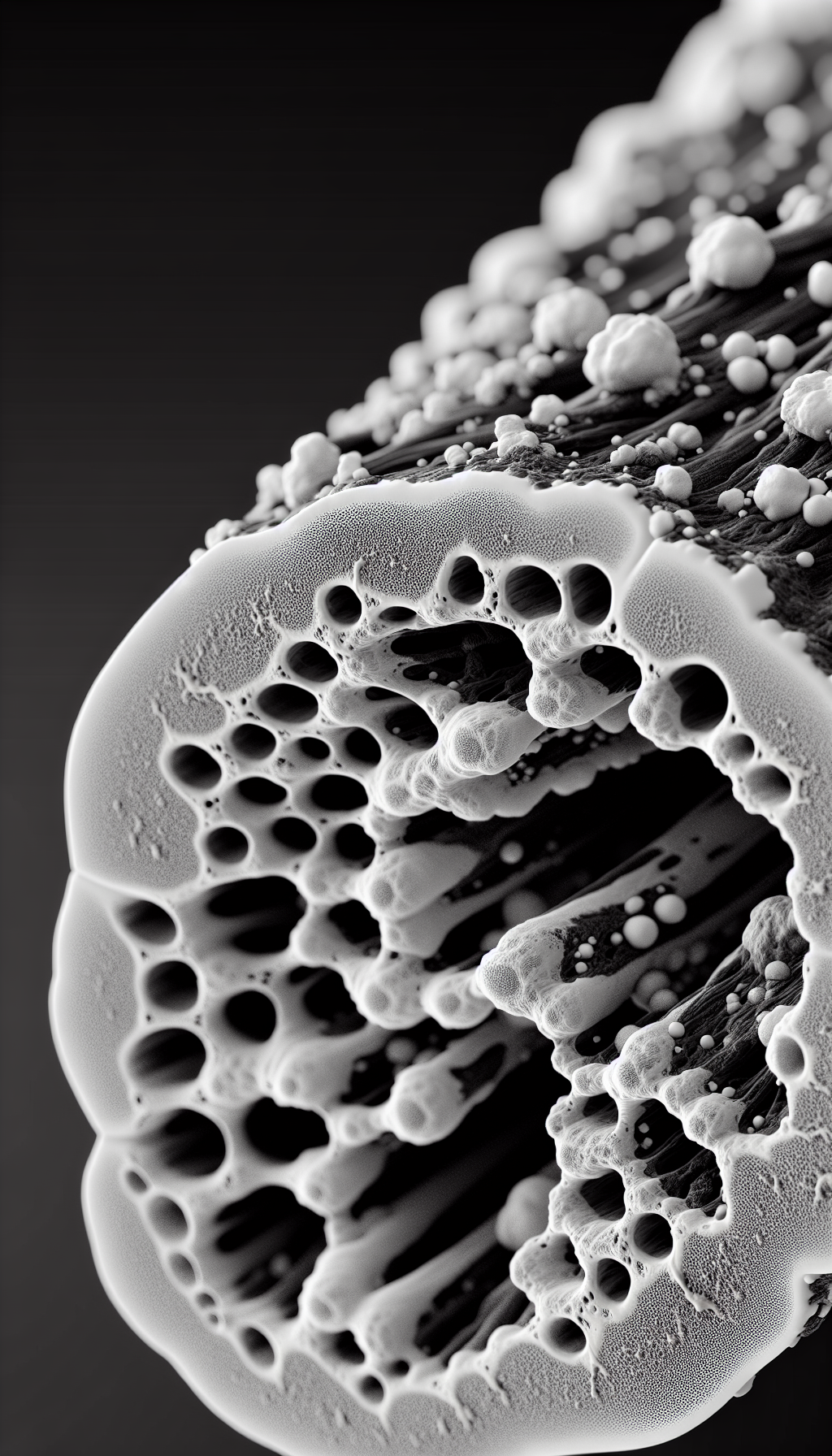
HR-pQCT offers a three-dimensional assessment of bone structure at a resolution capable of visualizing the trabecular lattice and cortical thickness. This high level of detail allows clinicians to better understand the microarchitecture of bone tissue, which is vital in predicting fracture risks beyond what BMD values can tell us.
Magnetic Resonance Imaging (MRI)
MRI, traditionally used for soft tissue imaging, has seen advancements that now allow for detailed bone analysis. Techniques like magnetic resonance micro-imaging (µMRI) can now provide high-resolution images of bone microstructures without exposing patients to radiation. MRI can also assess bone marrow composition, which has implications for overall bone health.
Quantitative Ultrasound (QUS)
Quantitative ultrasound is another imaging modality that’s gaining attention in the field of bone health. QUS devices measure the speed of sound through bone, which can reflect bone density and elasticity. As a portable and radiation-free option, QUS is an attractive tool for both clinical and field settings.
Implications of Advanced Imaging for Treatment and Prevention
The implications of these advanced imaging techniques extend far beyond diagnosis. They are reshaping the approach to treatment and prevention of bone-related diseases. For instance, by understanding the detailed architecture of bone, healthcare providers can tailor treatments more effectively.
Moreover, these imaging modalities can assess the impact of chronic inflammation on bone health, allowing for targeted interventions. Inflammatory conditions such as rheumatoid arthritis can lead to significant bone loss, and advanced imaging can play a crucial role in monitoring and managing these effects.
Integrating Lifestyle Interventions with Imaging Insights
In light of these imaging advancements, individuals can also take proactive steps to support their bone health. Diet and exercise, for example, have a profound impact on bone density. The benefits of yoga for bone health are well documented, and when combined with imaging data, practitioners can tailor programs that maximize bone strength and flexibility.
Nutrition is another cornerstone of bone health. A balanced diet rich in calcium and vitamin D is essential, but recent research also highlights the significance of silicon for bone health. Silicon plays a role in bone formation and maintenance, and knowing one’s bone density can inform dietary choices to include silicon-rich foods or supplements.
The Future of Bone Density Imaging
As we look towards the future, it’s clear that bone density imaging will continue to evolve. Research in the field is focusing on refining these technologies to make them more accessible and to enhance their predictive capabilities. Artificial intelligence and machine learning are also entering the fray, with algorithms being developed to analyze imaging data more precisely and to predict future bone health issues.
External Resources for Further Exploration
For readers interested in delving deeper into the subject, here are a few niche resources:
- The International Osteoporosis Foundation provides resources on bone health, including research on the latest imaging techniques.
- The National Osteoporosis Foundation’s Clinician’s Guide offers a detailed look at the clinical applications of bone density testing.
- BoneKEy Reports publishes peer-reviewed articles on bone research and the latest findings in bone health and disease.
The ongoing innovation in bone density imaging is a beacon of hope for those at risk of osteoporosis and other bone-related conditions. With continuous advancements, we can look forward to a future where these conditions are not only well-understood but also effectively prevented and managed.